International Medical University (IMU)’s Chinese Medicine programme integrates both western and Chinese Medicine subjects. One of the modules in Semester 6 is “Clinical Skills Development: Radiology” which helps students to master the ability to interpret the result of radiographic procedures such as X-ray, CT, MRI and more, to provide a basis for their diagnosis during clinical practice. Due to the restrictions by COVID-19 pandemic, students were unable to physically have their field visit to the Radiology Department in Hospital University Teknologi Mara (UiTM).
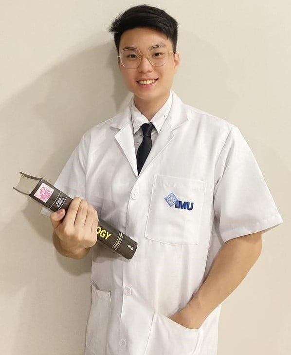
Hi, I am Chok Jun Yin, currently a Semester 6 Chinese Medicine student at IMU. I would like to share my valuable learning experience on a virtual field trip at the Radiology Department in Hospital UiTM on 1 September 2020. A virtual tour was conducted and we were pleased to have an experienced senior consultant radiologist, A/Prof Marymol Koshy to guide us in this session. Dr Marymol began the virtual tour with an introduction about the role of imaging modalities such as plain X-rays, computerized tomography (CT) scan, ultrasound scan and magnetic resonance imaging (MRI). She then demonstrated to us how to perform a chest X-ray and told us the importance of precautions taken for the patients, staff and relatives when performing a chest X-ray.
After that, the next destination was the control room. She also explained that X-ray operators will monitor their patients by observing through the viewing window to avoid any radiation exposure. If health care workers need to be present in the X-ray room, a lead apron must be worn to minimize radiation exposure. The X-ray technicians are required to wear dosimeters so a record of radiation exposure can be detected and measured for monitoring. She showed us some X-ray films and we were able to interpret the films correctly.
| Ultrasound Demonstration |
|---|
| Next, she invited her colleague to lie down on a bed. Then, she placed some gel on his abdomen to provide better contact between the body and the ultrasound probe. The two-dimensional pictures were shown instantly on a monitor. We were asked to describe the part of the organs shown in the monitor to familiarize with the internal structures of the body. Furthermore, Dr Marymol demonstrated the procedures for MRI examination and how to interpret the MRI images. She was pleased with our anatomy knowledge and our ability to answer most of the questions. |
Before ending the virtual field trip, we expressed our gratitude to Dr Marymol as it was not an easy task to guide us online. This was the first time that I experienced a virtual field trip, it was quite interesting to view the imaging modalities and learnt how to perform the imaging procedures. We were passionate and kept asking questions to gain a better understanding of our radiology study. I think a lifelong curiosity is an innate aspect that drives the learning progress and success. I felt bad that we could not pay a visit to Hospital UiTM because of this COVID-19 pandemic but I truly believe that we will get through this difficult time and visit the hospital again in the future.
This article is submitted by the Clinical and Experiential Learning Sub-committee.