8 November 1895 denoted one of the most important dates in the timeline of the history of medicine. It was the day when Wilhelm Conrad Röntgen, a German medicinal engineer and physicist, accidentally discovered a highly energetic electromagnetic radiation that he called X-rays, leading to a revolution in medical imaging. He was then honoured with the first Nobel Prize in Physics in recognition of his remarkable discovery.
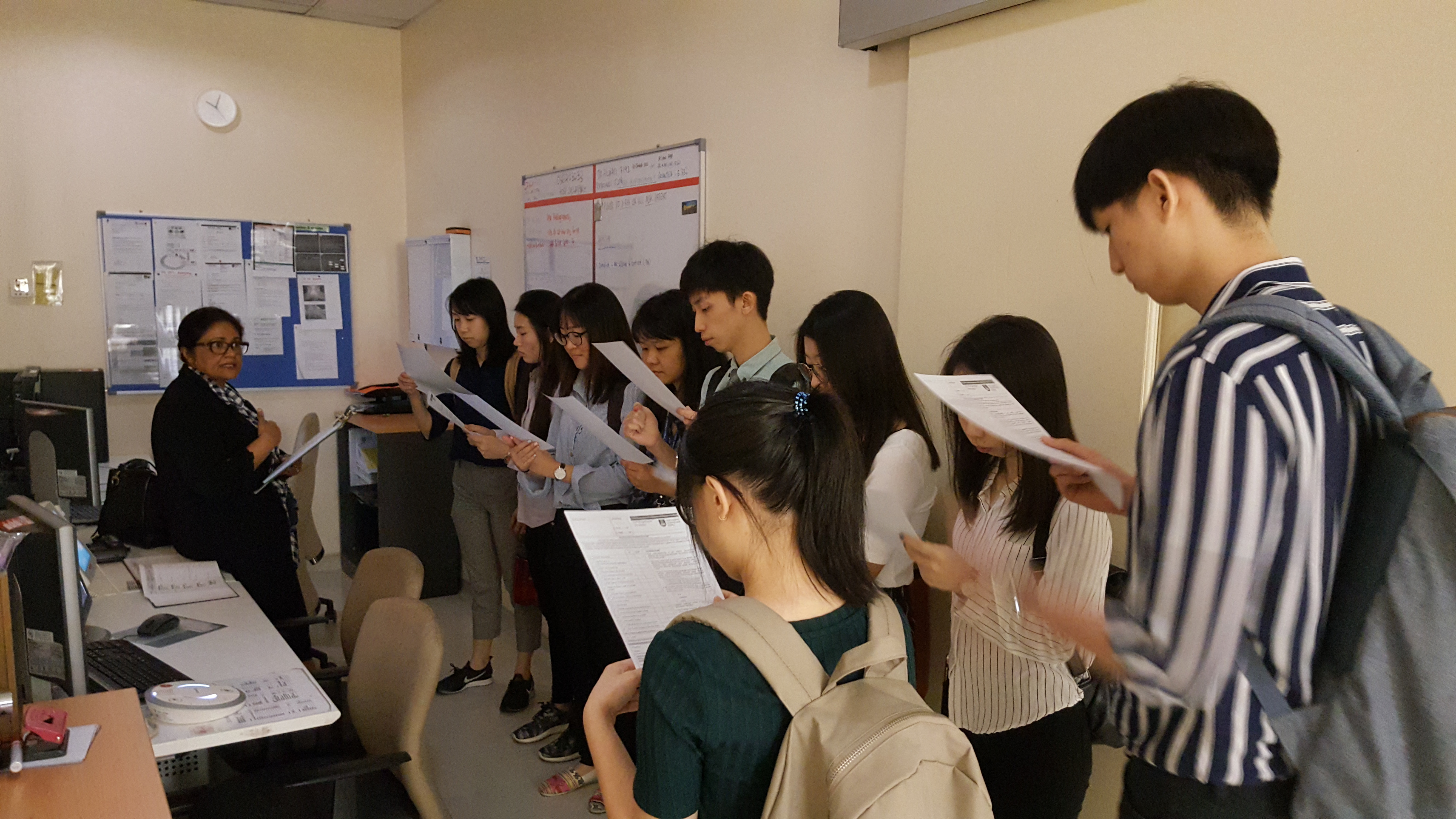
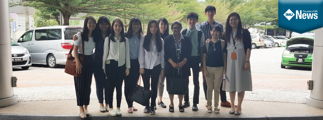
Recognising the importance of this, one of the modules in Semester 6 of the IMU Chinse Medicine programme (a unique semester that focuses on various Western Medicine modules) is dedicated to a branch of medicine that deals with X-rays and different imaging modalities – radiology. 26 August 2019- 9 IMU Chinese Medicine students (CM2/16) and a lecturer visited the Radiology Department at Universiti Teknologi MARA (UiTM), Sungai Buloh. The 2-hour visit was guided by an experienced senior consultant radiologist at UiTM, A/Prof Marymol Koshy. 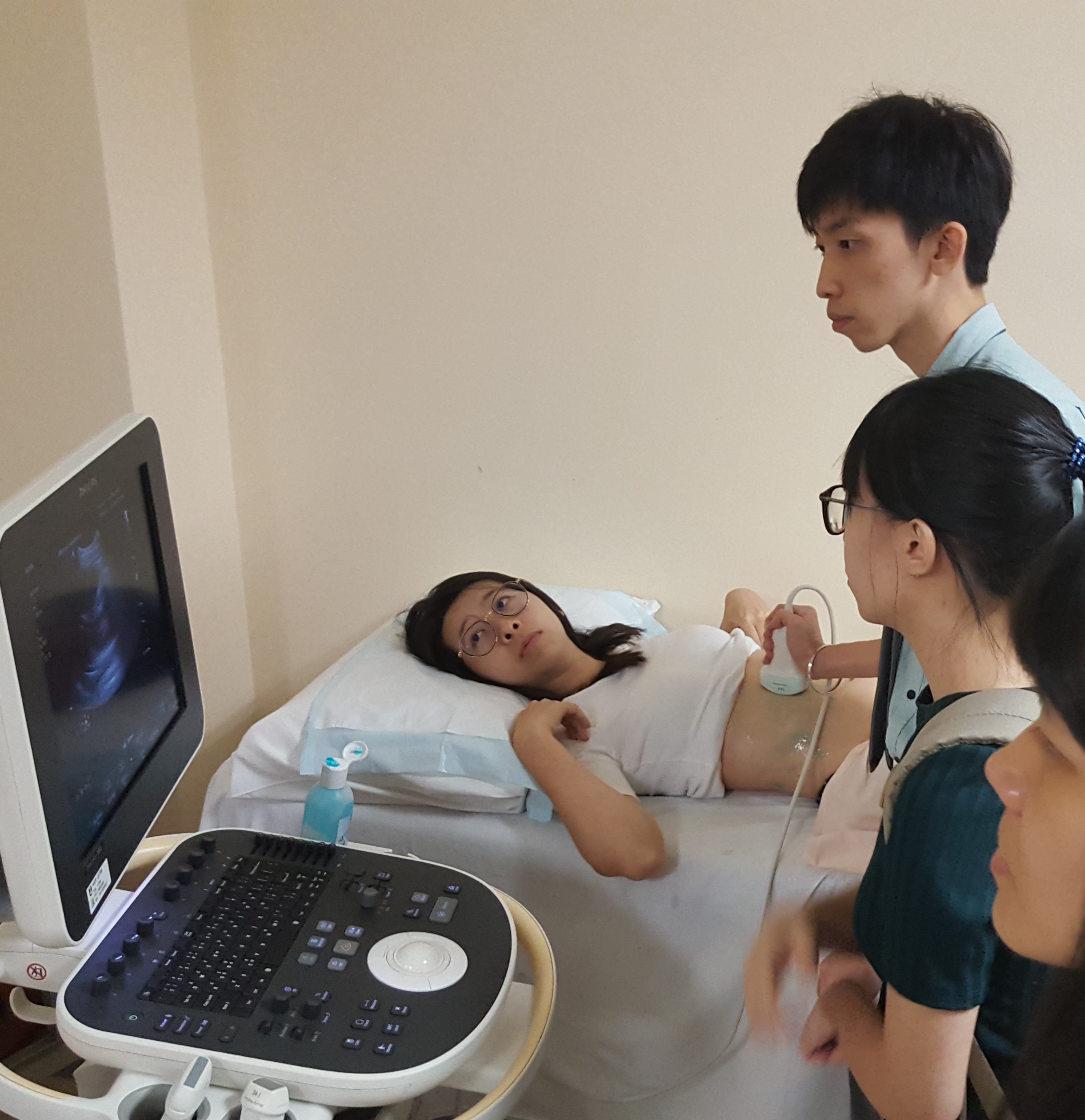 The visit began with a brief introduction to the general settings in a Radiology Department. Dr Marymol then shared the in-depth knowledge of different modern imaging modalities, including magnetic resonance imaging (MRI), computerized tomography (CT) scan, plain X-rays and ultrasound imaging. While visiting the MRI Department, Dr Marymol also demonstrated the pre-screening procedures before the MRI examination and the techniques to adjust, optimise as well as interpret some of the three-dimensional detailed MRI images in a computer. The students were attentive and able to answer most of the questions that Dr Marymol asked. Furthermore, Dr Marymol also explained about the pros and cons of a plain X-ray and ultrasound imaging. The students were allowed to use the ultrasound on each other and report the findings.
“Ultrasound imaging emits high-frequency sound waves and the image we see is the reflection of the sound waves. It is a useful tool to examine the fetus, gallbladder, liver, pancreas, spleen, kidneys and large blood vessels but not air-containing structures such as stomach and intestines,” stated Dr Marymol.
The visit ended with IMU students and lecturer expressing their gratitude to Dr Marymol for her generous invitation and guidance throughout the 2-hour visit. Written by Mr Desmond Tee Wei Kang (CM216).